
CHENNAI: Indian Institute of Technology Madras (IIT Madras) has released the world’s most detailed 3D Atlas of human brainstem, through its high throughput brain imaging and computing platform that transforms whole human brains into 3D cell-resolution atlases.
Developed by Sudha Gopalakrishnan Brain Centre (SGBC) at IIT Madras, ‘ANCHOR’ (Atlas of Neurochemical Characterisation of the human brainstem with 3D Reconstruction) comprises the most comprehensive, multi-modal, 3D maps and atlases of the human brainstem to date spanning from prenatal period to childhood and adult brains.
Congratulating the SGBC on their work, Prof V Kamakoti, Director, IIT Madras, “I always take pride that at IIT Madras we are exploring a lot of things but this particular exploration puts IIT Madras in the frontiers of the most complex creation that this world has witnessed – the human brain. This Centre is also studying brains affected by different diseases like rabies, dementia and Alzheimer’s disease. We now have a way by which we can say what happens to the basic structure of the brain due to diseases. This is a very important first step in understanding what happens in the human brain.”
These maps encompass more than 200 brainstem nuclei and fiber tracts, reconstructed from hundreds of serial sections. To resolve distinct neurochemical cell types, eight complementary immunotoxins were overlaid across more than 500 sections, enabling detailed mapping.
The atlas includes over 800 serial histological sections from human brainstems representing three stages of life—a 25-week gestational fetus, a 9-year-old child, and a 54-year-old adult—making it the most comprehensive human brainstem atlas developed to date. Researchers manually identified and annotated more than 200 brainstem structures across these specimens and characterized them using multiple neurochemical markers.
The platform also enables users to seamlessly navigate between MRI scans, block-face images, cellular-level tissue sections, and 3D reconstructions of the brainstem through an integrated online viewer, providing an unprecedented multi-scale view of this vital region of the human brain.
Prof. Mohanasankar Sivaprakasam, Head of SGBC, IIT Madras, added, “The key technology platform that makes these atlases widely accessible is our multi-modal image visualization framework that seamlessly integrates macro-scale volumetric data with micro-scale cellular images. By establishing precise spatial correspondence across these modalities, the atlas enables a seamless transition from gross brain structures in the MRI to cellular-level features. We envision that these maps and atlases will have significant implications for neuroscience and neuromedicine. This is an important scientific milestone for the Centre and is a big boost as we pursue our mission of imaging over 100 whole brains across the human lifespan and neurological diseases.”
SGBC aims to build the most comprehensive set of cell resolution human brain maps across life span and diseases. This Centre has become a truly global interdisciplinary team featuring more than 200 researchers, engineers and technicians working with 20 collaborators from different countries.
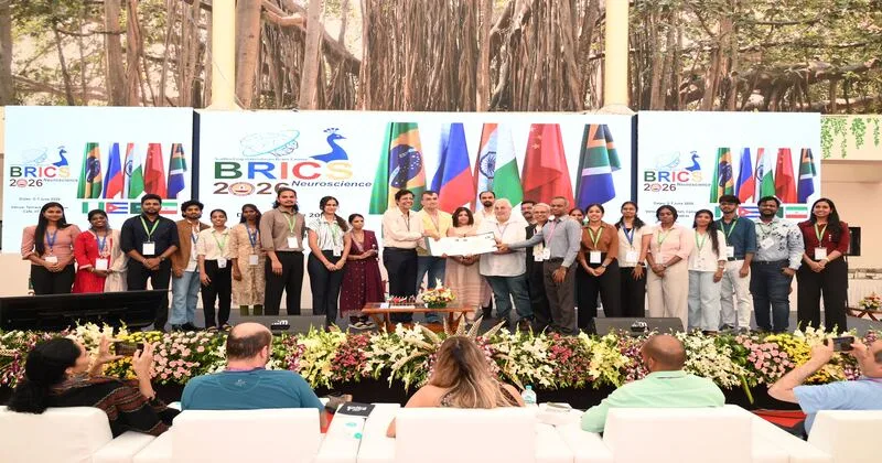
ANCHOR was released during the 3rd BRICS Neuroscience Symposium 2026 held from June 5-7 2026 at the IIT Madras campus. Institutions including CMC Vellore, Kilpauk Medical College, MediScan Systems, and Shri Ramachandra Institute of Higher Education and Research supported the Centre by facilitating the acquisition of brain specimens from individuals of different age groups and neurological profiles.
This collective effort enabled the creation of a diverse and comprehensive dataset, enriching the project’s scientific depth and helping advance a more representative understanding of the human brain.